Diagnostic procedures
When you go to the doctor for Achilles tendon pain, your doctor will examine your ankle and foot, looking for these signs:
- Swelling at the back of the heel and along your Achilles tendon
- Enlargement or thickening of your Achilles tendon
- Osteophytes or bony spurs on the lower part of the tendon at the back of the heel (insertional tendinitis)
- Where your Achilles tendon pain is the most intense
- Achilles tendon pain located in the middle portion of the tendon (non-insertional tendinitis)
- Pain located in the back part of the heel, at the lower section of your Achilles tendon (insertional tendinitis)
- Limited movement in your ankle. The doctor will specifically look for how well or if you can flex your foot
Sometimes imaging tests are ordered to help diagnose conditions related to Achilles tendon pain.
- X-RAYS. X-rays provide doctors with clear images of the bones. They can help show if the lower portion of the Achilles tendon has become hardened or calcified and is causing Achilles tendon pain. This hardening in the lower portion is an indication of insertional Achilles tendonitis. With severe non-insertional Achilles tendonitis, calcification can also be present in the middle section of the tendon.
- MAGNETIC RESONANCE IMAGING (MRI). Tendonitis as a cause for Achilles tendon pain can be diagnosed without an MRI, but this test is needed to plan for surgery to show how much damage has occurred in the tendon. The results of an MRI can help your surgeon determine what procedure is most appropriate to help correct the problem that is causing your Achilles tendon pain.
Conservative treatment
In the early stages of Achilles tendon pain, when there is acute or sudden pain and inflammation, one or a combination of the following conservative treatments might be recommended:
- Immobilization. This may include the use of a walking boot that can be removed or a cast to treat your Achilles tendon pain. Immobilization reduces the amount of force through the tendon and allows it to rest, promoting healing.
- Ice. To reduce Achilles tendon pain and to decrease swelling caused by inflammation, ice may be applied to the painful area for about 15 to 20 minutes every hour that you are awake. Use a thin towel between the ice and your skin. Never put ice directly on your skin.
- Oral medications. To relieve Achilles tendon pain, many doctors recommend nonsteroidal anti-inflammatory drugs (NSAIDs) because these medications also reduce inflammation. They include ibuprofen, aspirin, and naproxen.
- Orthotics. Some people have gait abnormalities such as over-pronation associated with Achilles tendon pain. In these cases, custom braces or orthotics may be ordered.
- Night splints. To help maintain the stretch in the Achilles while you sleep, a night splint may be recommended to decrease Achilles tendon pain.
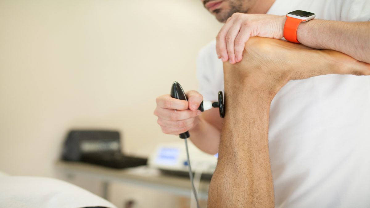
- Physical therapy. Physical therapy for Achilles tendon pain may include stretching, ultrasound therapy, strengthening exercises, running and gait re-education, and soft-tissue massage and mobilization.
- Cortisone injections. Corticosteroids are strong anti-inflammatory medications. They can be injected into the Achilles tendon, but they are rarely used due to the increased risk for tendon ruptures with these drugs.
Surgical Treatment
Surgery is typically only considered to relieve Achilles tendon pain if the pain does not resolve within six months of conservative treatment.
- Gastrocnemius recession. In this procedure, the calf muscles are made longer. This procedure benefits patients who remain unable to flex their feet or have pain with the movement, despite therapy to improve the condition. In gastrocnemius recession, one of the muscles of the calf muscles is lengthened to improve the mobility of the ankle.
- Debridement and repair. In this operation for Achilles tendon pain, when there is less than 50% damage to the tendon, the damaged section of the Achilles tendon is removed. After removal of the unhealthy tissues, the remaining portion of the tendon is repaired with stitches. If there are any bone spurs, they are also removed. Tendon repair in these cases may require the use of plastic or metal anchors to help anchor the tendon to the bone of the heel.
- Debridement with a tendon transfer. If greater than 50% of the tendon has been damaged, as is causing Achilles tendon pain, the remaining section would not be able to function. To prevent the healthy section from rupturing, a tendon transfer is completed. The tendon from the foot that assists in moving the big toe are moved to the bone of the heel bone. The big toe remains functional, and most patients do not notice any change in their gait.
After Surgery
Following surgery, the traditional method of treatment involves placing the surgical area in a brace or cast for up to eight weeks after the operation for the protection of the repair and incision. No weight-bearing is allowed, so crutches are needed at first.
Complications that may occur include scarring, delayed healing, and infection, as well as more serious complications like nerve damage and rupture of the tendon. To help avoid these complications, conditioning exercises done during this time help maintain aerobic fitness and adequate overall muscle strength of the patient.
Muscle wasting, blood clots, and joint stiffness can be complications of immobilizing the leg in a cast or brace. In order to prevent these problems, doctors might have patients start simple movement exercises very soon after their procedure. Patients typically wear a removable splint that they take off to perform the exercise during the day. A cane or crutch might be needed initially to help avoid limping.


Leave a Reply
You must be logged in to post a comment.