


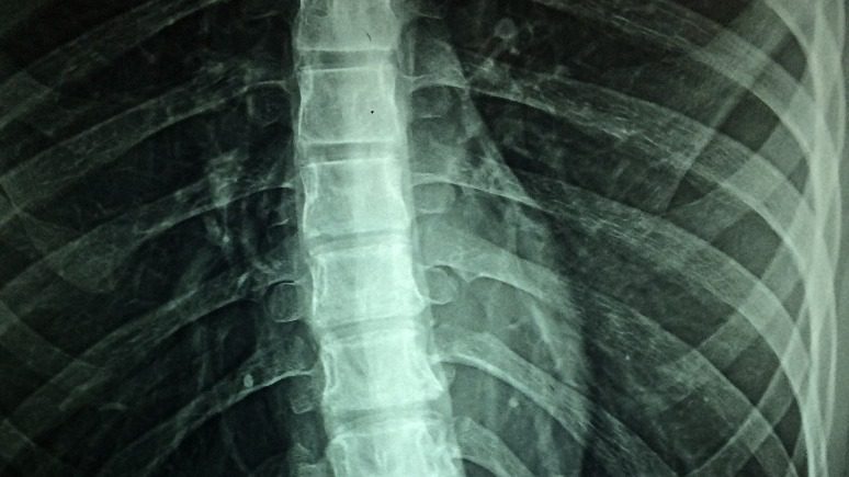

What is arthrography?
Arthrography, or an arthrogram, is a special kind of X-ray. It uses air or a contrast dye or sometimes both, along with a special x-ray to give your doctor a more detailed picture of the soft tissues of your joint. These tissues include muscles and cartilage, tendons, and ligaments. These tissues cannot be seen on plain X-rays.
The definition of arthrography includes two types of procedure: Direct and Indirect arthrography.
Direct arthrography is when the contrast material is injected by the radiologist into the joints directly. This imaging technique is usually more preferred than indirect arthrography because it enhancing the imaging of small internal structures, giving it a clearer and precise image. Direct arthrography helps improve the diagnosis and evaluation of the possible conditions and diseases of the joint.
On the other hand, indirect arthrography is when the contrast material is not injected into the joint directly but rather in the bloodstream. The contrast material will then eventually be absorbed by the joint.
The Purpose of Arthrography
This procedure is commonly used to determine what causes joint pain and discomfort. In addition, local anesthetic medications or steroids are sometimes injected together with the contrast material so that any inflammation or joint-related pain is decreased. These medications help physicians to receive further information on what might be causing the joint pain.
The reasons your doctor might recommend an arthrography are:
- To find out what is causing unexplained and chronic problems in your joints, such as swelling, pain, or unusual movement
- To look for signs of tears, degeneration, or disease inside the joint or in the cartilage or ligaments
- To look for disturbances in the way the bones meet and join each other
- To look for abnormal cysts or tumors
- To guide needle placement when fluid is taken from the joint for analysis
An arthrogram can be completed on several joints such as the jaw, wrist, elbow, shoulder, ankle, knee, or hip. Sometimes an MRI or a CT scan is used with arthrography to give the doctor more information about what is going on in a joint.
If your doctor recommends arthrography, be sure to inform him and the technician performing the procedure if you:
- Have any drug allergies, including allergies to anesthetics
- Have an allergy to iodine. Iodine may be used in the contrast material
- Have an allergy to shellfish or bee stings
- Might be or are pregnant
- Have rheumatoid arthritis
- Have asthma
- Are taking blood thinners or have a bleeding problem
- Have an infection in your joint or near it. The contrast material could make the infection worse
Preparation and Procedure
Preparing for the procedure can vary depending on the method your doctor will use.
Before the arthrogram begins, you will be asked to remove any metal objects or jewelry from around the joint being studied. You will then lie or sit down with your joint positioned under a special X-ray machine called a fluoroscope connected to a monitor. The area will be thoroughly cleaned and covered, and you will be injected with a local anesthetic to help make you comfortable during the procedure.
A needle will be inserted into the joint, and some fluid may be extracted to make more room for the contrast material to be injected. Some of the fluid that is removed may be sent to the lab to be analyzed. The contrast material is then injected, and as it enters the joint, you may experience pressure, pain, tingling, or fullness in the joint.
You may be asked to walk around or move in different positions to help the contrast material become evenly distributed. Other than when you are asked to move, you should stay as still as possible. It is important for the x-rays to be taken quickly before the contrast spreads to the other tissues outside the joint. Typically it takes 30 minutes to one hour to complete the test.
After the Procedure
Following arthrography, you will need to rest the joint for at least 12 hours. Then, avoid exertion for one or two days. Using ice packs can help keep any swelling that may occur to a minimum. Over-the-counter pain medications like acetaminophen or aspirin are usually effective for pain management after arthrography.
Results of the Procedure
The results of the arthrography are interpreted by radiologists who are trained specifically to understand and interpret any radiology examinations. After the images are available, the radiologist will send a report to your referring physician, who will be the one to discuss and relay the results of the examination with you.
In some cases, it may be necessary to undergo follow-up examinations. Your referring physician will explain why this is requested. Follow-up examinations are done if a potential abnormality needs to be further evaluated. Most of the time, follow-up examinations are needed to monitor any known abnormality in order to see best if your treatment is working or not.


Leave a Reply
You must be logged in to post a comment.